


The canine spine (vertebral column) is not only the supporting structure of the body, it also carries the spinal cord, which is composed of nerves that move muscles, originate sensations, and act as a connecting link to all the systems of the body. Preserving the health of the spine begins with a physically fit dog. It is especially important to keep the dog at the proper weight to avoid added stress on this all-important part of the dog’s anatomy. I stress to my puppy buyers that allowing their dog to become overweight is essentially “killing them with kindness” and should be avoided at all cost. Simply put, overfeeding your dog leads to an early grave.
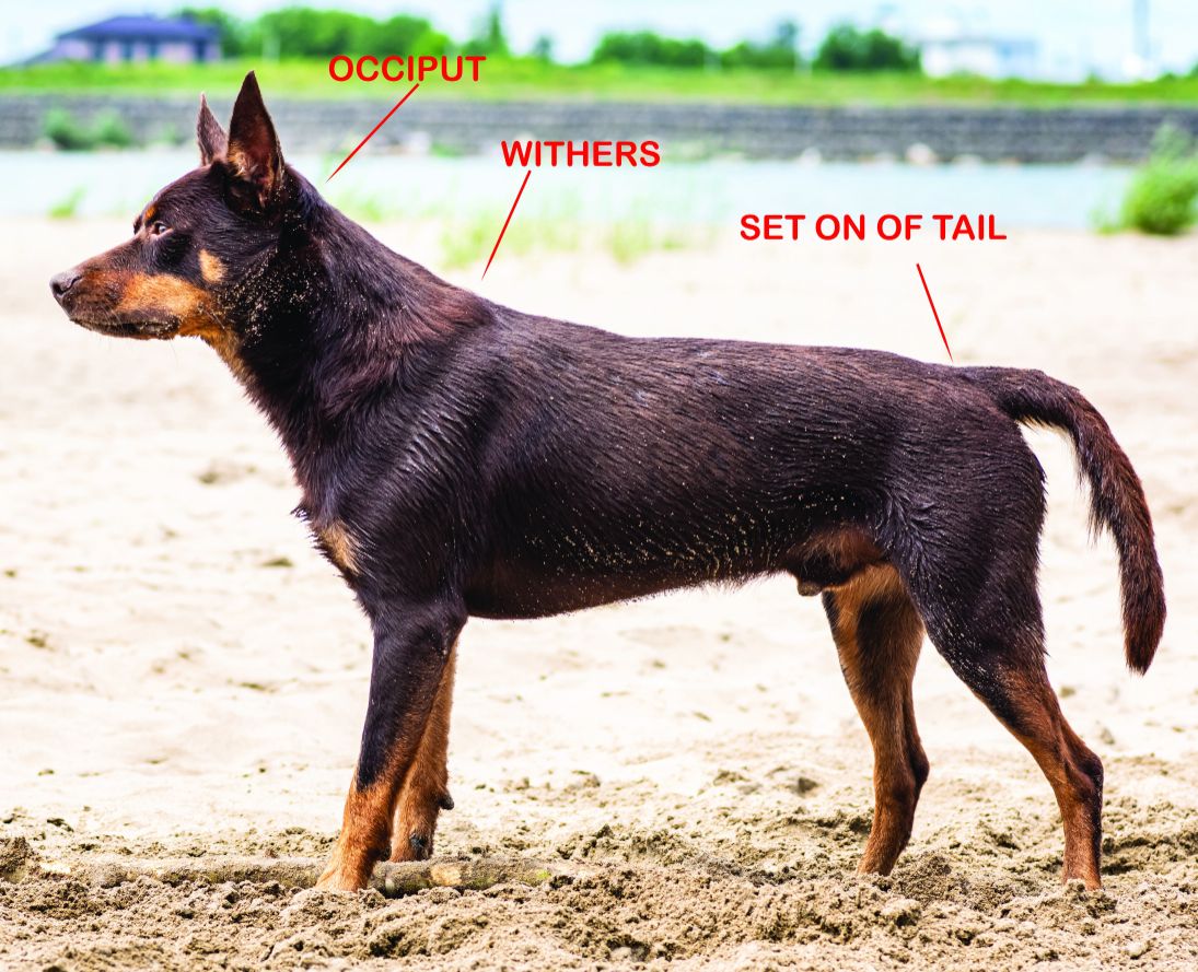
General terminology used when discussing the spinal column as a whole is the “topline’” of the dog. The topline begins behind the ears at the occiput, proceeds over the neck, withers, back, and croup, ending at the set-on of the tail. In some breed standards, the term is used to mean the “backline,” which is only a section of the topline from the withers to the base of the tail.
The canine spine functions to support the head, fore and hindquarters, and rib cage, and to support and protect the internal organs. This column of vertebrae allows for movement of the head and back of the animal. The encasing and protection of the spinal cord, which transmits messages to and from the brain and throughout the body, is a major function of the spinal column. The canine spine also serves a vital part in transmitting the force generated by the hind limbs throughout the rest of the body in order to propel the dog forward.
A quick overall assessment of the canine spine will tell us that the spinal column is made up of multiple bones (50 in number) called vertebra, which form a sort of chain through which the individual vertebra move against one another. There are five sections of the vertebral column; cervical (neck), thoracic (chest), lumbar (loin), sacrum and coccygeal (tail). Each vertebra has its own identifying number. Cervical are numbered C1-C7, Thoracic are T1-T13, Lumbar are L1-L7, and Sacral are S1-S5. The coccygeal vertebrae vary in number from none (no tail) to anywhere from 20-25. Most breeds average 23 coccygeal vertebrae, designated by using Cd and a number.

While many a standard may call for a dead level topline, there is a curvature of the spine that can be seen in Figure 2 and is demonstrated by the dashed line. Because of the length of coat and the musculature that is overlaid onto the canine spine, as well as the varying lengths of the spinous processes, the topline can appear to be flat. Again, this can vary from breed to breed, so one must be familiar with the standard that is specific to their breed of dog. On physical examination, the curves are a bit more evident. However, even when placing your hands on the dog, it may still be difficult to feel the curves due to the condition of the thoracic vertebrae at the first thoracic spine; run your forefinger or thumb down the middle of the neck toward the backline until you can feel a bump in the pathway, somewhere (hopefully) between the tips of the shoulder blades.

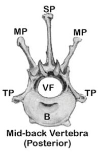
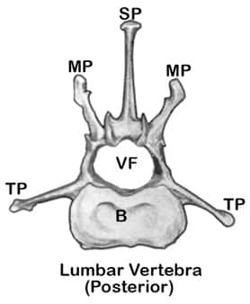
The largest part of the vertebra is the body (B). There are several smaller projections from the central part of the bone, with smaller surfaces that articulate with similar projections on neighboring vertebrae. Projecting from the top of the vertebra is the top spine (SP – Spinous Process), which functions as a base for muscular attachment. The shape and size of the spinous process varies in the different regions of the canine spine, according to the amount of muscle attachment needed, and can vary (sometimes widely) from breed to breed. More spinous projections for muscle attachment are found on either side of the vertebra. The transverse processes (TP) are situated at somewhat right angles to the long axis and the mammillary processes (MP) extending from side-to-side. The large hole in the vertebra (VF) is the vertebral opening (foramen) through which the spinal cord passes.
The shape and size of the individual vertebra are determined by the function of that section of the spinal column. We will separate the spinal column into four basic sections; the neck (cervical), the chest (thoracic), and the loin (lumbar), plus the sacrum and the tail (caudal). It is relatively easy to see the differences in the shape of these three vertebrae, from the first thoracic vertebra to the thoracic vertebra, from the mid-back, and then from the lumbar section. (See Figures 3, 4 & 5.)


The cervical vertebrae support the head and allow for its movement up-and-down and from side-to-side as well as enable the dog to extend the neck forward or toward the ground.
Between the vertebrae are intervertebral discs that are composed of a compressible substance and are attached to each vertebra, allowing for movement and serving as a shock absorber. During my research, I think the most interesting description of these discs was comparing them to jelly donuts—with the dough being the fibrous covering on the outside of the disc and the inside being the jelly or gelatinous material (nucleus pulposis) serving as the cushioning inside the disc. (See Figure 6.) As a dog ages, the jelly inside the disc becomes chalky and harder. When the disc is damaged due to deterioration or injury, it can leak the jelly-like or hardened interior, which is termed a herniated disc. As the interior is extruded, it compresses the soft tissues and nerves surrounding the disc, causing pain and immobility. When the discs degenerate, the condition is referred to as intervertebral disc disease or IVDD.
The Neck (Cervical Vertebrae)
The canine neck contains seven cervical vertebrae as do all mammals, with only a few exceptions. Think on this for a moment: Mammals, from humans to giraffes, have seven cervical vertebrae. The only difference is in the length of the cervical bones. The cervical vertebrae support the head and allow for its movement up-and-down and from side-to-side as well as enable the dog to extend the neck forward or toward the ground.
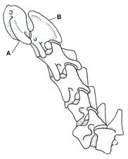
The first two vertebrae of the neck are unique in their formation and function. The first cervical vertebra is the atlas (“A” in Figure 7), which allows up-and-down movement (think “yes” movement of the head), and the second is the axis (“B” in Figure 7), which allows the side-to-side or rotary movement associated with the “no” movement of the head. The Atlas has a smaller body and spinous process, but also has very long, thick transverse processes called “wings of alae,” which are easy to feel in the neck. The wings allow for the attachment of powerful neck muscles from which arises the much-admired arch in the neck of a dog. The atlas attaches to the skull in a unique way and allows for the hinge-like up-and-down movement of the skull on the neck. Most of the cervical vertebrae do not have the high spinous process as shown in the thoracic vertebrae in Figures 3-5. The neck vertebrae tend to be wider in body, with spinous processes that gradually increase in length to the last (7th) vertebra. In some of the larger breeds, the cervical vertebrae are malformed, leading to disc rupture and compression of the spinal cord. This is commonly referred to as “Wobblers Syndrome.”
The Thoracic Vertebrae
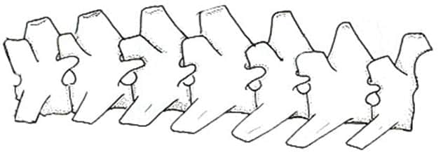
There are 13 thoracic (chest) vertebrae. Their primary function is the support of and attachment to the ribs for the vital process of breathing. The longer spinous processes allow for the attachment of the strong neck and back muscles. The first nine thoracic vertebrae have the longest spinous processes, and they all angle back toward the tail of the dog. These vertebrae comprise what is called the “withers” of the dog (most especially the second and third vertebrae, which are the highest). Even more important, the first nine ribs are attached to both the thoracic vertebrae at the top and to the sternum underneath the dog, forming a nearly solid, enclosed unit. The last four thoracic vertebrae, which have tips that are fairly level with one another, form the mid-back of the dog. Some refer to this section as the “true” back, as these four vertebrae are nearly identical in shape to the lumbar vertebrae, but they still fully function as rib-carrying thoracic vertebrae. (See Figure 8.)
The lumbar area and the last thoracic vertebrae play a more important role in movement when the dog is at a gallop.
The Lumbar Vertebrae
The seven lumbar vertebrae support the organs of the abdomen and the pelvis, and offer protection of the abdominal organs and the spinal cord. They are vital in the movement of the dog. The loin area of the canine spine rises upward in a curve that then slopes downward toward the pelvis and then on to the three sacral vertebrae and the first several tail (coccygeal) vertebrae that form the croup. The lumbar vertebrae gradually increase in width from the first to the seventh, with bodies that are longer and heavier than the thoracic vertebrae, and increase in length from the first through the sixth. The last lumbar vertebra is shorter than those that precede it, making it about the same length as the first lumbar vertebra. In the average dog, the first nine thoracic vertebrae are about equal in length to the seven lumbar vertebrae. The spinous and transverse processes of the lumbar vertebrae are massive, to allow for strong muscle and ligament attachments that are vital for the transmission of power from the rear of the dog. (See Figure 9.) Because of the shape of the lumbar area and the position of the articular facets of the individual lumbar vertebrae, free flexion and extension and a slight rotation are allowed, though this inhibits much lateral movement. Because of this special formation of the lumbar canine spine, it is not unusual for a dog to show a slight roach over the lumbar area that is temporary and does not affect the dog in motion. The lumbar area and the last thoracic vertebrae play a more important role in movement when the dog is at a gallop. They begin to “roach” as the dog brings both rear legs forward under the body, which causes more forward reach, and then the same vertebrae begin to straighten out, increasing the rear extension and adding power to the force of forward propulsion through the powerful back muscles. The reach of the spinal cord ends at the fourth lumbar vertebra.
Sacral Vertebrae and Croup
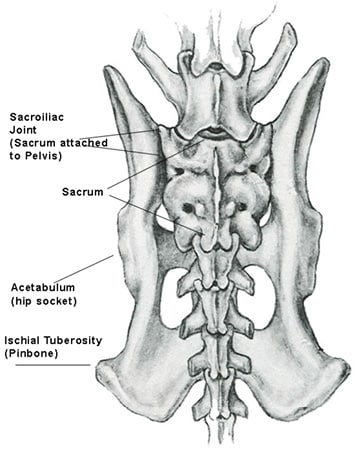
The sacral vertebrae consist of three vertebrae that are fused together. (See Figure 10.) The important function of the sacral vertebrae is that on either side, it is joined by a cartilaginous joint with the pelvis. This forms a firm union that allows the transfer of forces from the hindquarters to the vertebral column. It is extremely important to understand that the hind limbs are attached to the pelvis with a ball and socket joint, and that the pelvis is firmly attached to the canine spine via the sacral
vertebrae, whereas the shoulder blade is attached to the body with only muscles and ligaments. This is one of the main reasons that we can see much more lateral movement in the forequarters than we can see in the rear, as there is a less rigid connection to the body in the forequarters (so there is space for “wiggle” room).
The croup is that area between the rim of the pelvis, which includes the fused sacral vertebrae, and the first four or five of the tail vertebrae. These vertebrae form a slightly curved area that can be fairly easy to feel through a physical examination on a dog in correct weight (not fat)! The angulation of the croup determines tail set. A fairly level croup indicates a higher tail set, and a more angled croup indicates a lower tail set. It is important to note that the angulation of the croup and the angulation of the pelvis are two separate and distinctly different parts of the body, and are independent of each other. You can have a dog with a flat croup and a flat pelvis, and one with a steep croup and a steep pelvis. But you can also have a flat croup and a steep pelvis as well as a steep croup and a flat pelvis.
The final set of vertebrae are the tail (coccygeal) vertebrae that can number from no tail at all through a full tail of approximately 20-23 vertebrae.
If you have any questions or comments, or would like to schedule a seminar, you may contact me via email: jimanie@welshcorgi.com.