


Canine Joints, Let us do a brief review of some of the basics.
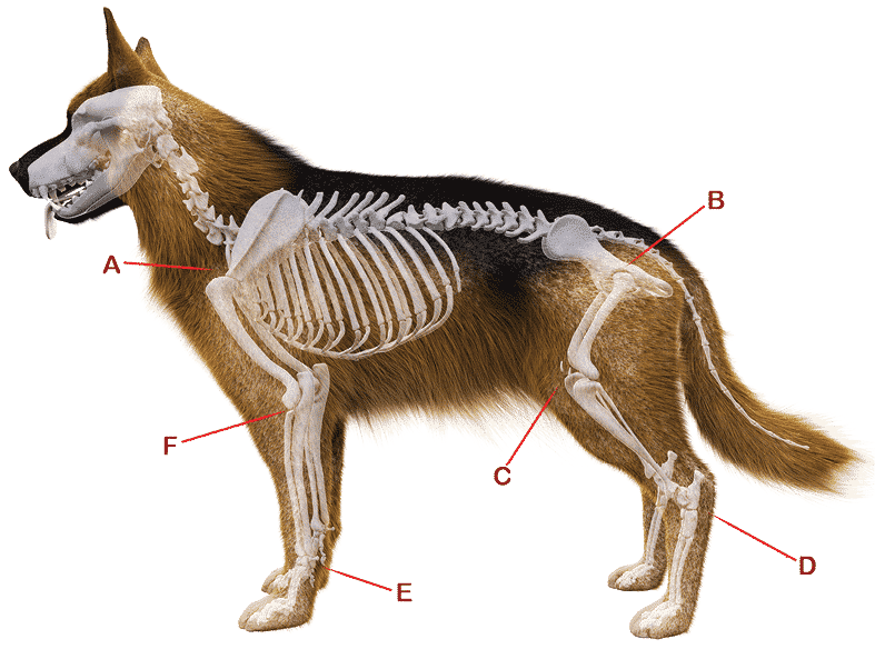
ANGULATION refers to the angles created by bones meeting at various joints (articulations), especially at the shoulder (Fig. 1A) and pelvic areas (Fig. 1B) and the stifle (Fig. 1C), hock (Fig. 1D), pastern (Fig. 1E) and at the elbow (Fig. 1F).
BONE comprises the structure of the skeletal system and provides lever arms for locomotion. Bone also plays important roles in maintaining mineral homeostasis (the balance of minerals, especially calcium and phosphorus). The bones must supply sufficient area for the attachment of the muscles. Smooth muscles account for one-third to one-half of the total body weight.

MUSCLES move the bones and dictate where they will go and where they will stop. The skeleton is simply bone, and has no means of creating motion by itself. Although we are concentrating on the inner structure of the skeleton and its articulations and relationship, without muscles there is no movement. The musculature of the dog is a complicated subject, too lengthy for us to approach here. We are simply going to try to understand the structural limitations imposed by the composition of the juncture of the various bones, and leave the study of the muscles of the dog up to the individual student.


TENDONS connect muscle to bone and are part of the muscle structure.
LIGAMENTS are NOT part of the muscle structure. They are tough, fibrous bands developed at joints to connect bones to other bones. Other ligaments form slings to hold tendons at the shoulder, wrist, and ankle joints.
CANINE JOINTS
There are “joints” (as in popular “joints” where people meet up), and then there are the joints formed within the body. Basically, a joint or articulation is a point where two or more bones meet and are united by fibrous, elastic or cartilaginous tissue or a combination of these tissues. There are three main types of joints: Fibrous (immovable); Cartilaginous (partially moveable); and Synovial (freely moveable).
Fibrous Canine Joints are those that are stationery, such as the joints in the skull, which allow little to no movement. The bones are held together tightly by tough, fibrous, connective tissue. (See Figure 2.)
The canine skull is actually made up of over 40 different bones, all tightly held together by this fibrous tissue. These various fibrous joints, also called “sutures” (see Figure 3A) serve to allow enough movement to absorb the shock of a blow as well as allow for growth at the edges of the bone. The fibrous joints range from those where a slight degree of compressibility is advantageous to those where a more extreme stability is desirable. A fibrous joint may have a considerable amount of intervening connective tissue. (See Figure 3.)
Cartilaginous Canine Joints allow some movement as compared to a fibrous joint, but less movement than a synovial joint. Cartilaginous joints are formed when two or more bones are joined together entirely by cartilage. The joints formed between each vertebra in the spine are cartilaginous joints, allowing a smooth, frictionless movement of the spine. (See Figure 4.) The intervertebral disc is composed of fibrocartilage, which joins two vertebrae together. The center of the disc consists of a gel-like material. Another example of cartilaginous joints are the joints where the ribs meet the sternum.
Synovial Joints are, by far, the most common classification of a joint within the canine body.
They are highly moveable and all have a synovial capsule (collagenous structure) surrounding the entire joint, a synovial membrane (the inner layer of the capsule) that secretes synovial fluid (a lubricating liquid), and cartilage known as hyaline cartilage that pads the ends of the articulating bones (fibrous tissue enclosing a synovial cavity). The joints are lubricated for smooth action by synovial fluid and are stabilized by tendons and ligaments. The joints are the hinges of the body. A well-formed joint allows bones to act as levers; the bones move at angles to each other to produce movement. Internally, they have cushion-like padding called cartilage. Externally, they are held together by flexible ligaments.
There are six types of synovial joints, which are classified by the shape of the joint and the movement available.
The dog’s body has three basic types of joints that we are going to discuss:

left to right: Figure 5. Upper Arm/Shoulder Blade Joint; Figure 6. Pelvis/Femur Hip Joint; Figure 7. Elbow Joint; Figure 8. Stifle Joint
A BALL AND SOCKET JOINT moves in two directions with rotational capabilities. It is formed by a convex hemispherical head, which fits into a cavity. (See Figures 5 & 6 and Figure 9 for an x-ray of a hip joint.)
A HINGED JOINT moves on one axis (like a door). It permits flexion and extension with a limited degree of rotation. The most moveable surface of a hinged joint is usually concave. (See
Figures 7 & 8.)
In the GLIDING or PLANE JOINT, the articular surfaces are nearly flat. The bones that make up the joint can glide or rotate. The pastern joint (carpus joint) of the dog is located approximately in the same position as the human wrist. (But it is NOT a wrist!) The carpal bones in this joint form two rows. (See Figure 9.) It is a synovial joint, comprised of a common outer fibrous capsule and three inner synovial pouches, one for each joint. Numerous ligaments add to the stability of the joint and ensure that movement is largely limited to a gliding motion and a very small amount of rotation.
(See Figure 9A.) The pastern serves as the dog’s main shock absorber and allows for flexibility in movement. Let me repeat myself: Pasterns absorb the impact of every step ever taken by your dog. (See Figure 9.)
A CONDYLAR JOINT resembles a hinged joint in movement, but involves a prominence in one bone fitting into a depression in the articulating bone. A good example is the stifle. (See Figures 10, 10A & 10B.) It looks much like a knuckle, and results in two articular surfaces, usually included in one articular capsule. These knuckle-shaped condyles vary in distance from one another, allowing uniaxial movement with limited rotation.
After my dogs had their way with a cow bone, I saved it to photograph as it really does demonstrate how a condyle joint fits together. (Figure 11 shows the leg bone and the carpal bones separately. Figure 12 shows how well they fit together.)
Canine Joints | If you have any questions or comments, or to schedule a seminar, contact Stephanie via email: jimanie@welshcorgi.com or by using SSM contact form.

left to right: Figure 9. Pastern (Carpus) Joint; Figure 9A. Pastern (Carpus) Joint; Figure 10. Stifle Condyles
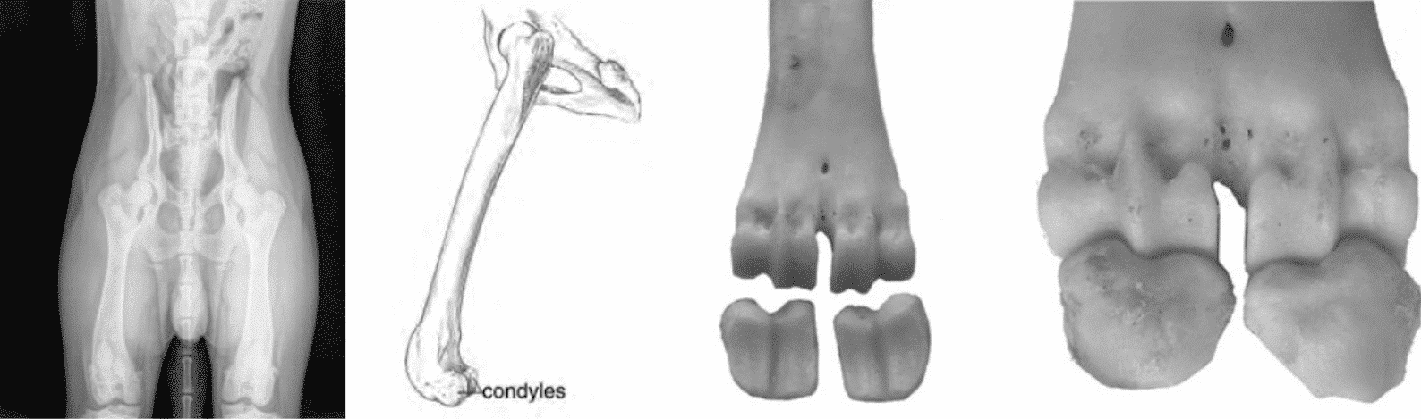
left to right: Figure 10A. Hip Ball and Socket Joint and Condyles on Femur (Young German Shepherd Dog); Figure 10B. Condyles on Femur; Figure 11. Separated Condyle Joint; Figure 12. Condyle Joint Articulation