


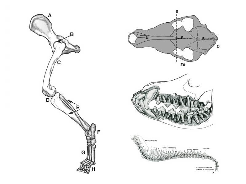
Today, we will deal with the structure of the hindquarters and a bit about the head, neck, and spinal column. This should give us a basic understanding of the skeletal structure of the dog, which we can then relate to the shape and movement of the dog in the following installments. As a reminder of how all the parts fit together, see Figure A.
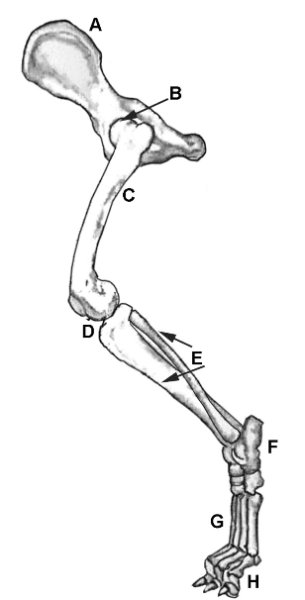
The hindquarters of the dog are comprised of the pelvis (A), hip joint (B), femur (thigh or upper thigh) (C), stifle (knee joint) (D), leg, which may be referred to as the second or lower thigh (tibia – a larger bone and fibula – a smaller bone) (E), hock joint or ankle joint (tarsals) (F), rear pastern (metatarsals) (G), and the bones of the foot containing the foot joint and the toes (phalanges) (H).
These bones are fused together to form the pelvis. In most breeds, the pelvis is approximately the same length as the shoulder blade (scapula). The pelvis usually lies at a 30 degree angle to the horizontal. (See Figure 2).
The hip joint forms an angle of approximately 90 degrees between the pelvis and the femur. The hip joint (B) is of major importance in the structural support of the dog and is at the center of the mechanical force that provides for locomotion. The upper thigh (femur) (C) is the major bone of the rear leg and is approximately the same length as the pelvis. The knee (stifle) (D) is the joint between the upper thigh and second thigh (fibula and tibia) (E) (fib-tib for brevity).
The fib and tibia are the bones that make up the leg and are equal to or somewhat longer than the pelvis and femur. The tibia is the main weight-bearing bone of the leg. The fibula provides lateral stability to the ankle joint. The fib-tib may be referred to as the ‘second or lower thigh, but it actually corresponds to the leg in the human.

The tarsal bones form the hock joint (F) (ankle) and form an approximate angle of 120 degrees between the fib-tib and the metatarsals. The rear pastern (metatarsals) (G) are the bones of the foot. These bones are located between the hock joint and the dog’s foot and are commonly included when referring to the “hock” of the dog. Technically, the “hock” only refers to the joint, but its usage is so common that it is widely understood and, therefore, accepted when describing the metatarsals or rear pastern. The metatarsals (G) form an approximate 90-degree angle with the rear toes (phalanges) (H) of the dog.
The hock joint (ankle) (F) consists of seven bones arranged in two rows. The hinge joint formed by the articulation of the upper tarsal bones with the tibia and fibula prevents lateral shifting of the joint. The fibular tarsal bone is the large, long bone that is the insertion point for the calcaneus tendon (Achilles tendon in humans), from which a strong leverage effect is possible. The top of the fibular tarsal bone, which sticks up from the joint at the top of the hock, is the calcaneal process (spur bone), and it is to this projection that the calcaneus tendon is attached.
In a breed such as the Whippet, you can often see the delineation of the tendon, especially in strong daylight. A closer examination of this joint is fascinating in the way the bones fit together and articulate with each other in order to offer the highest amount of stability possible to the joint. The metatarsals, or rear pastern (G), are the bones of the foot (remember that a dog walks on his toes).
These bones are located between the hock joint and the dog’s foot and are commonly included when referring to the “hock” of the dog. Technically, the “hock” only refers to the joint, but its usage is so common that it is widely understood and, therefore, accepted when describing the metatarsals or rear pastern. The bones of the metatarsus are similar to the metacarpus of the front foot, with the exception of being longer than those of the metacarpus. The metatarsals (G) form an approximate 90-degree angle with the phalanges (H), or rear toes, of the dog.
Three different types of heads are found in the dog. The average head is termed mesocephalic; “cephalic” from the Greek refers to the head, and meso basically means “medium” and refers to a square head. This is the most common term pertaining to the overall head. The more proper term for the ratio of skull to muzzle is “mesaticephalic”. This average head has a closer balance of muzzle to skull length as in a German Shepherd Dog or Miniature Pinscher.
The head with a shorter muzzle and wider head is termed brachiocephalic (brachio meaning short) such as the Bulldog or English Toy Spaniel, and finally, the long, narrow head is a dolicocephalic type of head (dolico meaning long) such as the Collie or the Borzoi. Most breeds have a mesocephalic or mesaticephalic shaped head. The areas of importance mentioned in most standards are the zygomatic arch (ZA) (cheek bones), the area of the stop (S), the length and width of the muzzle (nasal bones N), the length and width of the skull (F, the forehead or frontal bones, and B, the brain case or backskull), and the occipital protuberance (O), which is the back point of the skull.

The parts of the skull are mostly self-explanatory. The skull in Figure 3 represents the average medium (mesocephalic) head of our average dog. There is a great deal of variance between many breeds in the form of the skull. Each breed has a head unique to that breed, just as each breed has a gait unique to that breed. Some of the dogs developed for the same set of tasks are very similar in head shape, structure, and gait, but there are always exceptions to the rules!
The brain case, or back part of the skull (B), encloses and protects the brain. Even though the head varies greatly from breed to breed, the size of the brain remains about the same relative to the type of skull and the size of the dog. Therefore, a long-headed dog has the same brain capacity as the smaller, short-headed breed of dog. The frontal bones (F) are roughly the same as the human forehead and are the bridge between the backskull or brain case and the muzzle or nasal bones (N).
The stop (S) is the indentation between the eyes and is the separation point between the top planes of the muzzle and the skull. It is part of the frontal bone (F). The position of the stop in the skull and the angulation where the top of the muzzle meets the skull vary from breed to breed and are important breed characteristics no matter the breed.
The depth of the stop is dependent upon the size of the sinus cavity (air sac) formed in the frontal bones of the skull to lessen the weight of the head. A large, rounded sinus forms a deeper stop than an elongated, narrow cavity in a breed that calls for a slight or nearly no stop. Sinus cavities allow for voice resonance, help filter and add moisture to the air inhaled through the nasal passages, limit the weight of the skull, help to insulate and cool the brain, and allow increased insertion space for teeth.
The muzzle is composed of the nasal bones that form the top plane of the muzzle as well as the maxillary bones, which form the base for the teeth in the upper jaw, and the mandible, which forms the lower jaw. At this point, I would interject that in any breed that interacts with livestock, vermin, or game birds or animals, complete dentition should be considered the ‘norm’ for those breeds. Especially dogs that deal with livestock and may take a kick to the jaw. See Figures 4 and 5.
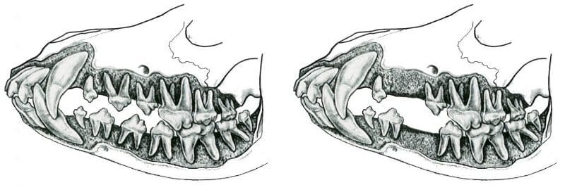
The roots of the teeth are a strengthening and stabilizing factor for the jaws, especially in the lower jaw. A forceful kick that lands on an area of the mandible that lacks a large tooth (whether a molar or premolar) is much more likely to break than if the dog had full dentition. Just one more thing for you to ponder.
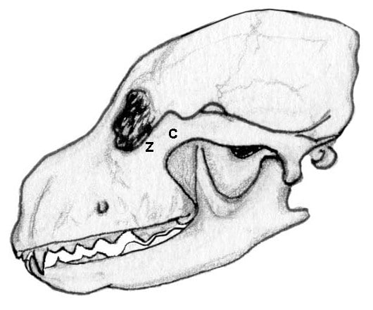
The zygomatic arch (Z, Figure 6) is an important factor in the shape of the skull. A small band of bone, it forms part of the bottom of the eye socket and sweeps backward in an arch, the degree of which differs from breed to breed. The average dog has a moderately curved zygomatic arch, whereas the shorter-skulled dog has a much wider, more pronounced curve.
Conversely, the longer, narrower-headed dog has a correspondingly long, narrow, flat zygomatic arch. The back portion of the arch (C, Figure 6) also forms the cheek bones, and its thickness is a contributing factor to the coarseness of the head of the dog.
The dog’s bite is another important consideration in the formation of the skull. The bite will depend upon the relative length of the lower jaw as compared to the length of the upper jaw. There are so many facets to the head of the dog that are vital components that make up the overall breed type that I could spend hours on the head alone. Since this is about the basics, we will now leave the head and discuss the neck and the rest of the spinal column.
The spine of the dog is the connecting link between all of the “parts” we have discussed in the previous installments of this series. The spine functions to support the head, fore and hindquarters, the rib cage, and protect the internal organs. This column of vertebrae allows for movement of the head and back of the animal. The encasing and protection of the spinal cord is a major function, as is its vital part in transmitting the force generated by the hind limbs throughout the rest of the body in order to propel the dog forward.
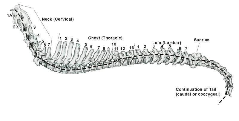
A quick overall assessment of the spine will tell us that the spinal column is made up of multiple bones called vertebra, which form a sort of chain in which the individual vertebra move against one another.
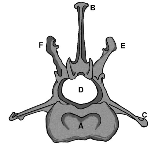
The largest part of the vertebra is the body (A). There are several smaller projections from the central part of the bone with smaller surfaces that articulate with similar projections on neighboring vertebrae called laminae (layers) or facets (flat surfaces). Projecting from the top of the vertebra is the top spine (B) (Spinous Process), which functions as a base for muscular attachment.
The shape and size of the spinous process vary in the different regattachment, asions of the spine according to the amount of muscle attachment needed. Two more spinous projections for muscle attachment are found on eithevertebra andf the vertebra are called the transverse (extending from side to side; situated at right angles to the long axis rocess (C).
The large hole (D) is the vert(foramen),ning (foramen) through which the spina cord passes. Other log projections such as E and F also allow muscle attachment and the smaller projections and facets, which, depending upon the direction in which they face, allow for either sliding lateral or sideways movement or up and down movement, but allow for very little rotary movement in the spine.
Again, these facets and laminae allow for specific movement depending upon the section of the spine in which they occur. Between the vertebrae are intervertebral discs, which are composed of a compressible substance and attached to each vertebra, allowing for movement and serving as a shock absorber.
While the Standard may call for a dead-level topline, there is a curvature of the spine, which can be seen in Figure 7 and is demonstrated by the dashed line. Because of the length of the coat and the musculature that is overlaid onto the spine, as well as the varying lengths of the spinous processes, the topline can appear to be flat.
On physical examination, the curves are a bit more evident, though even when placing your hands on the dog, it may still be difficult to feel the curves. Just about everyone can palpate the end of the neck and the beginning of the chest at the first thoracic spine by running their forefinger or thumb down the middle of the neck toward the backline until they can feel a bump in the pathway somewhere between the tips of the shoulder blades.
The shape and size of the individual vertebra are determined by the function of that section of the spinal column.
Whenever a speaker begins to talk about the canine neck, they usually start with the joke: “Short neck or long neck, all dogs have the same neck.”
Since the neck contains seven cervical vertebrae, this is a true statement. With only a couple of exceptions, all mammals, from humans to giraffes, have seven cervical vertebrae! The only difference is the length of the cervical bones. The cervical vertebrae both support the head and allow for its movement up and down and from side to side, as well as enabling the dog to extend the neck forward or toward the ground.
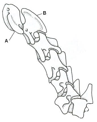
The first two vertebrae of the neck are unique in their formation and function. The first cervical vertebra is the atlas (A in Figure 10), which allows up and down movement (think “yes” movement of the head), and the second is the axis (B in Figure 10), which allows the side-to-side or rotary movement associated with the “no” movement of the head.
The Atlas has a smaller body and spinous process but very long, thick transverse processes called “wings of alae” which are easy to feel in the neck. The wings allow for the attachment of powerful neck muscles, from which arises the much-admired arch in the neck of a dog. The atlas attaches to the skull in a unique way and allows for the hinge-like up and down movement of the skull on the neck.
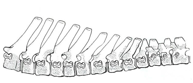
Most of the cervical vertebrae do not have the high spinous process as shown in Figure 8. The neck vertebrae tend to be wider in the body, with spinous processes that gradually increase in length to the last (7th) vertebra.
There are 13 thoracic (chest) vertebrae, and their primary function is the support and attachment of the ribs for the vital process of breathing. The longer spinous processes allow for the attachment of the strong neck and back muscles. The first nine thoracic vertebrae have the longest spinous processes and all angle back toward the tail of the dog. These vertebrae comprise what is called the withers of the dog, especially the second and third vertebrae, which are the highest.
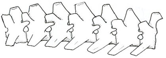
Even more important, the first nine ribs are attached to both the thoracic vertebrae at the top and to the sternum underneath the dog, forming a nearly solid, enclosed unit. The last four thoracic vertebrae, which have tips that are fairly level with one another, form the mid-back of the dog. Some refer to this section as the ‘true’ back, as these four vertebrae are nearly identical in shape to the lumbar vertebrae but still fully function as rib-carrying thoracic vertebrae.
The seven lumbar vertebrae support the organs of the abdomen and the pelvis, as well as offering protection to the abdominal organs and the spinal cord. They are vital to the movement of the dog.
The loin area of the spine rises upward in a curve that then slopes downward toward the pelvis and then on to the three sacral vertebrae and the first several tail (coccygeal) vertebrae that form the croup. The lumbar vertebrae gradually increase in width from the first to the seventh, with bodies that are longer and heavier than the thoracic vertebrae and increase in length from the first through the sixth. The last lumbar vertebra is shorter than those that precede it, making it about the same length as the first lumbar vertebra.
In the average dog, the first nine thoracic vertebrae are about equal in length to the seven lumbar vertebrae. The spinous and transverse processes of the lumbar vertebrae are massive to allow for strong muscle and ligament attachments, vital for the transmission of power from the rear of the dog. Because of the shape of the lumbar area and the position of the articular facets of the individual lumbar vertebrae free flexion and extension and a slight rotation is allowed, but it inhibits much lateral movement. Because of this special formation of the lumbar spine, it is not unusual for a dog to show a slight roach over the lumbar area that is temporary and does not affect the dog in motion.
The lumbar area and the last thoracic vertebrae play a more important role in movement when the dog is at a gallop. They begin to roach as the dog brings both rear legs forward under the body, which causes more forward reach, and then the same vertebrae begin to straighten out, increasing the rear extension and adding power to the force of forward propulsion through the powerful back muscles. The reach of the spinal cord ends at the fourth lumbar vertebra.
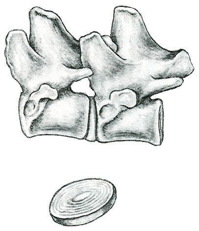
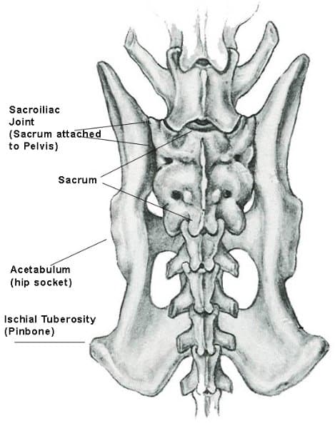
The sacral vertebrae consist of three vertebrae that are fused together. (See Figure 11). The important function of the sacral vertebrae is that on either side, they are joined by a cartilaginous joint with the pelvis. This forms a firm union that allows the transfer of forces from the hindquarters to the vertebral column. This is extremely important to understand that the hind limbs are attached to the pelvis with a ball and socket joint, and the pelvis is firmly attached to the spine via the sacral vertebrae, whereas the shoulder blade is attached to the body with only muscles and ligaments. This is one of the main reasons that we can see much more lateral movement in the forequarters than we can in the rear, as there is a less rigid connection to the body (so space for ‘wiggle’ room).
The croup is that area between the rim of the pelvis that includes the fused sacral vertebrae and the first four or five of the tail vertebrae. These vertebrae form a slightly curved area, which can be fairly easy to feel on physical examination of a dog of the correct weight (not fat!). The angulation of the croup determines tail set. A fairly level croup indicates a higher tail set, and a more angled croup indicates a lower tail set.
It is important to note that the angulation of the croup and the angulation of the pelvis are two separate and distinctly different parts of the body that are independent of each other. You can have a dog with a flat croup and a flat pelvis, and also one with a steep croup and a steep pelvis, but you can also have a flat croup and a steep pelvis, as well as a steep croup and a flat pelvis.
The final set of vertebrae are the tail (coccygeal) vertebrae, which can range from no tail at all to a full tail of approximately 20 vertebrae.
I have presented the very basics of structure. For those still awake, in the next installments I will try to make sense of the actions of this structure when the dog is in motion. As always, questions and comments are welcome via email at jimanie@welshcorgi.com.
Form Follows Function: Part Three. From the March 2019 Issue of Showsight Magazine.
This series originally appeared in the Working/Herding Digest. All rights reserved by the author.